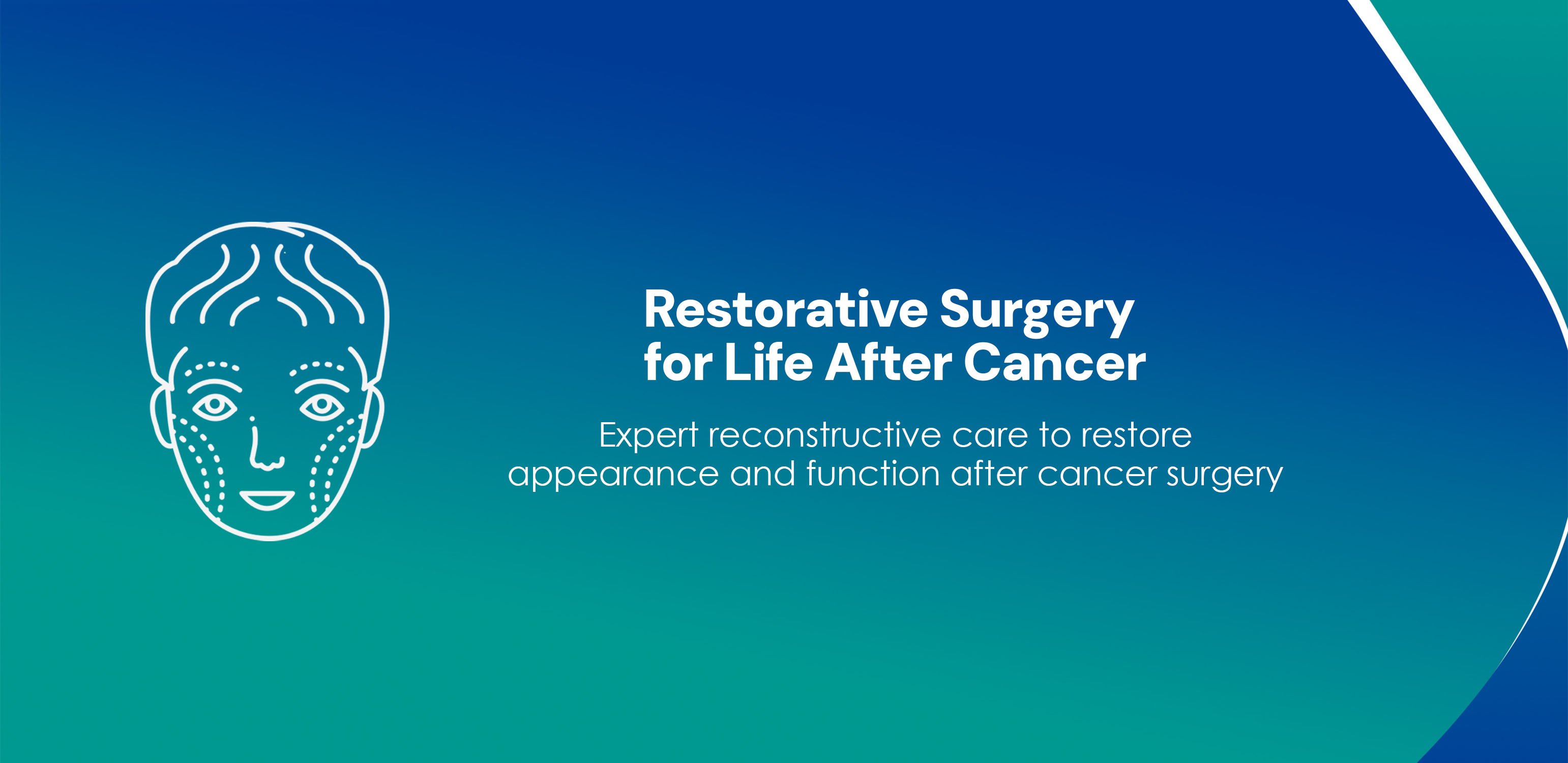
Restoring Form, Function, and Confidence After Cancer Surgery
When cancer is removed, surgeons sometimes need to take out skin, fat, muscle, bone, or other tissues to make sure all the tumor is gone. While this is lifesaving, it can leave defects that change how someone looks and functions—affecting speech, swallowing, breathing, movement, or body image.
Plastic & Reconstructive Surgery rebuilds these areas using healthy tissue taken from elsewhere on the patient's body (called flaps). These flaps carry their own blood supply and help the wound heal strongly and reliably. Reconstruction may be done at the same time as tumor surgery (immediate reconstruction) or later (delayed reconstruction) depending on cancer treatment plans.
Head & neck cancers can involve the tongue, mouth, jaw, throat, cheek, and face. These areas are essential for speech, swallowing, chewing, breathing, and expression.
Taken from: Outer part of the lower leg (fibula bone), often with overlying skin.
Placed at: Jaw (mandible) when segment of jaw is removed.
Why used: Fibula provides a strong bone segment that can be shaped to make a new jaw. Dental implants can often be placed later.
Donor-site expectation: Most people walk normally; leg is stable. Expect a scar on the lower leg and possible numbness or weakness for a short time.
Taken from: Inner forearm skin and thin soft tissue (may require a skin graft to close the donor site).
Placed at: Tongue, oral lining, floor of mouth, or small cheek defects.
Why used: Thin, flexible tissue ideal for lining and mobile areas like the tongue.
Donor-site expectation: Forearm scar; temporary sensitivity change; a small graft area that needs care.
Taken from: Outer thigh skin and fat (sometimes muscle perforators).
Placed at: Large cheek, neck, or face soft-tissue defects.
Why used: Provides bulk when needed, good long scar hidden by clothing.
Donor-site expectation: Thigh scar, usually well hidden; walking is typically unaffected.
Taken from: Upper back/shoulder blade area.
Placed at: Composite defects needing both skin and soft tissue.
Donor-site expectation: Back scar; usually minimal functional loss.
Taken from: Chest muscle (pectoralis major).
Placed at: Large or emergency neck/cheek defects when quick, robust coverage is needed.
Donor-site expectation: Chest scar; small impact on pushing/weight-lifting but daily activities usually preserved.
Breast reconstruction restores breast shape after lumpectomy or mastectomy. It addresses physical balance as well as emotional and psychological well-being.
Taken from: Lower abdomen (skin and fat, sparing the muscle).
Placed at: Chest to create a breast mound.
Why used: Gives a natural-feeling breast using your own tissue; spare muscle means less donor-site weakness.
Donor-site expectation: Tummy scar similar to a "tummy-tuck"; improved abdominal contour.
Taken from: Lower abdomen including muscle.
Placed at: Chest.
Why used: Good tissue volume but may weaken abdominal wall more than DIEP.
Donor-site expectation: Tummy scar; possible abdominal weakness risk (surgeon may use mesh to reinforce).
Taken from: Upper back muscle and skin.
Placed at: Chest (often combined with implant for volume).
Why used: Reliable blood supply, useful especially when prior radiation has affected chest tissues.
Donor-site expectation: Back scar hidden by bra; small effect on shoulder strength for some patients.
Taken from: Synthetic implant (silicone or saline).
Placed at: Chest to recreate breast shape.
Why used: Shorter operation, quicker recovery; may be combined with flaps if needed.
Donor-site expectation: No donor scar; implants may need replacement over many years.
Restores breast shape and symmetry.
Improves clothing fit, posture, and body image.
Supports emotional and psychological recovery after cancer.
Children's bodies are still growing, so reconstruction requires special planning to preserve growth and function.
Protects bones and nerves after tumor removal.
Restores normal function and appearance as the child grows.
Reduces long-term deformities and improves quality of life.
Sarcomas often require removing large amounts of muscle and skin, exposing bone and joints. Reconstruction protects these structures and helps save the limb.
Taken from: Thigh skin/fat.
Used at: Limb or trunk defects—very versatile.
Taken from: Upper back.
Used at: Large defects especially in shoulder/back region.
Taken from: Inner thigh muscle.
Used at: Smaller limb defects or for functional muscle transfer.
Taken from: Leg bone.
Used at: When bone replacement is required (e.g., large tibial/mandible defects).
Taken from: Tissue close to the defect when available.
Used at: Moderate-size wounds to minimize new donor sites.
Saves limbs in many cases.
Restores movement, weight-bearing, and daily activities.
Reduces infection and wound breakdown, allowing chemotherapy/radiation to continue on schedule.
Radiation therapy is often part of cancer treatment. Radiation affects skin and soft tissue by making them thinner, firmer, and slower to heal. This influences reconstruction planning.
Better long-term appearance and function.
Lower risk of wound breakdown during radiation.
Faster overall recovery and return to normal life.
Wound healing and pain control; limited mobility.
Increasing activity with physiotherapy; many return to daily light activities.
Significant recovery; strength and function improving.
Long-term results apparent, scar maturation continues.
Your surgeon will discuss risks specific to your condition and comorbidities before surgery.
Yes — one at the cancer site and one at the donor site. Both heal and become less visible over time.
Yes — reconstruction restores shape and symmetry. Final results improve over months.
Yes — speech/swallow therapy or limb physiotherapy is often needed for best recovery.
Yes — but timing and flap choice are planned carefully with oncologists.
Yes. Modern implants are safe; some may need replacement in 10–15 years.
Sensation may be reduced initially but often improves over time.
Most daily activities resume in 4–6 weeks. Full healing & scar softening: 6–12 months.
Most patients return to normal activity. Fibula donors walk normally. Forearm donors regain hand function. Thigh donors walk normally. Back flap donors regain shoulder strength.
No — it is functional and reconstructive. It restores appearance and important everyday functions.
Reconstruction is a vital part of cancer treatment — it helps rebuild form and function, protects vital tissues, and improves emotional recovery.
Flaps are your own healthy tissues moved from a safe donor site to rebuild the area that cancer surgery removed.
Choice and timing matter — your surgical team will recommend the best flap and timing based on tumor type, need for radiation, and your personal goals.
Recovery needs time and therapy, but outcomes are often excellent: patients regain function, appearance, and confidence.
Ask questions and bring a support person to appointments — understanding the plan helps you feel more confident and prepared.
MBBS, MS - General Surgery (2015), MCh - Plastic and Reconstructive Surgery (2018)
ConsultantPlastic and Reconstructive Surgery
MBBS, MS - General Surgery (May 2005), DNB - General Surgery (Jan 2006), MRCS (Apr 2007), M.Ch - Plastic & Reconstructive Surgery (Aug 2008), Diploma in Medico Law & Ethics (Aug 2007)
Visiting ConsultantPlastics and Reconstructive Surgery
MBBS, DNB Plastic Surgery (2019)
Visiting ConsultantPlastics and Reconstructive Surgery