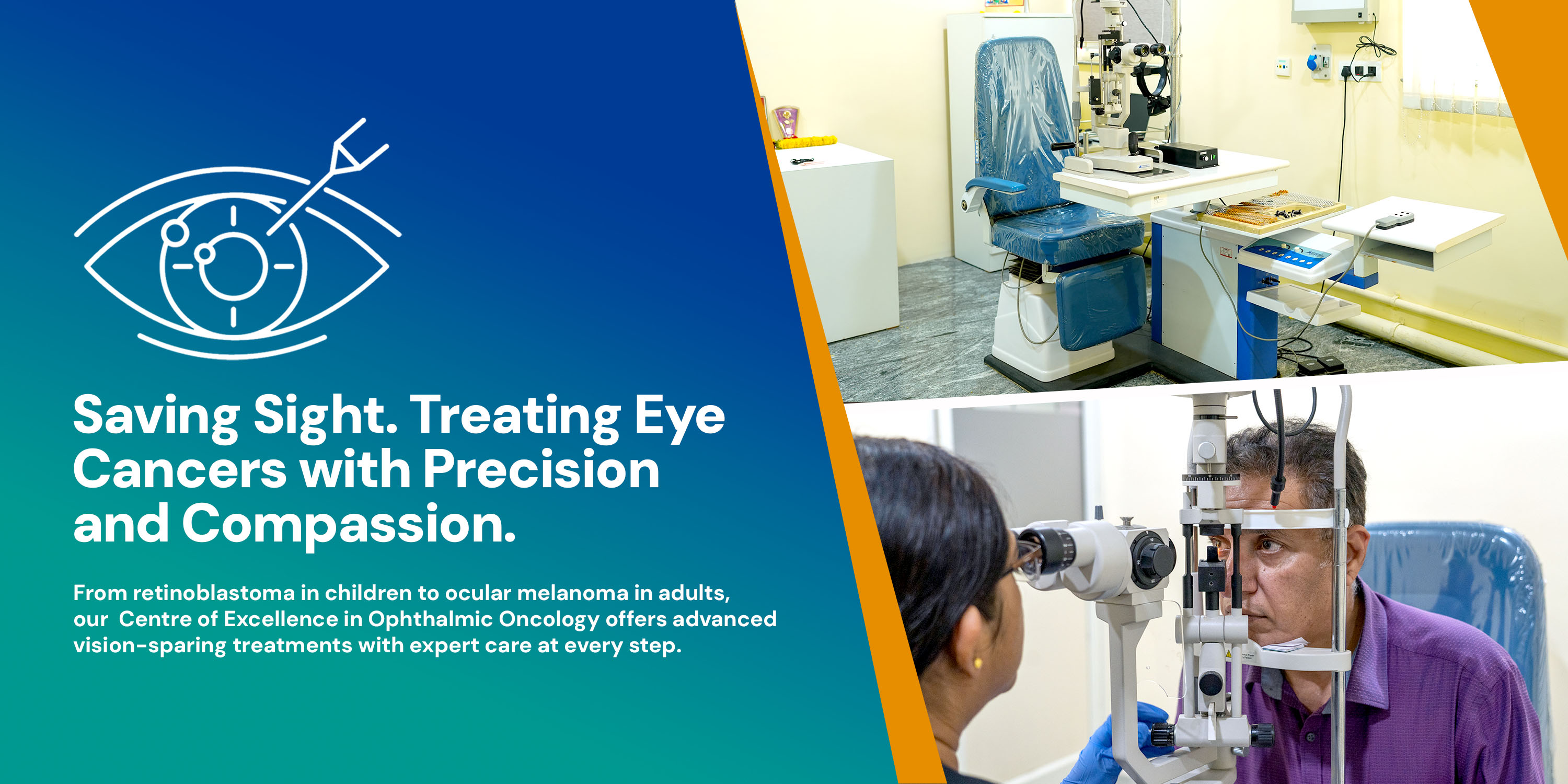
Eye cancer, or ocular malignancy, encompasses a diverse group of cancers that can develop within the eye or in structures surrounding it. While rare compared to other malignancies, they represent serious threats to both vision and life. Modern advances in diagnostic imaging, chemotherapy delivery, radiation techniques, and surgical innovation have dramatically transformed treatment, enabling vision preservation in the majority of cases while maintaining excellent survival outcomes.
Eye cancers range from highly curable intraocular malignancies in children to more aggressive systemic cancers in adults. Early detection and specialized, multidisciplinary care are critical to achieving the best functional and survival outcomes.
Retinoblastoma is the most common eye cancer in children, occurring in approximately 1 in 20,000 births worldwide. It develops in the developing retina (the light-sensitive tissue at the back of the eye) and accounts for 2–3% of all childhood cancers. The disease can occur in one eye (unilateral, ~60% of cases) or both eyes (bilateral, ~40% of cases), with bilateral cases always representing hereditary disease.
Outcomes: Retinoblastoma represents one of the oncological success stories: with contemporary globe-sparing treatment strategies, eye salvage rates exceed 75–95% (depending on disease stage), and overall survival rates exceed 95% in developed countries.
Uveal melanoma is the most common intraocular malignancy in adults, arising from melanocytes (pigmented cells) in the iris, ciliary body, or choroid (the uveal tract)—the middle pigmented layer of the eye. Unlike cutaneous melanoma, uveal melanoma is not related to sun exposure and occurs sporadically.
Landmark clinical trials have demonstrated that enucleation (eye removal) and brachytherapy (radioactive plaque therapy) result in equivalent long-term mortality rates, establishing that globe-sparing approaches are appropriate for appropriately selected tumors. Today, brachytherapy is the preferred globe-sparing treatment, allowing preservation of vision while achieving excellent disease control.
Ocular Surface Squamous Neoplasia (OSSN) encompasses a spectrum of dysplastic and malignant lesions affecting the conjunctiva and cornea. Risk factors include chronic sun exposure, human papillomavirus (HPV) infection, and immunosuppression. Treatment has evolved from exclusively surgical excision to include topical pharmacotherapy, reducing recurrence rates and preserving ocular surface health. Other conjunctival cancers include conjunctival melanoma and rare lymphomas affecting the ocular surface.
Orbital lymphomas represent a diverse group of lymphoid malignancies affecting the space surrounding the eye. The majority are B-cell lymphomas, ranging from indolent (low-grade) to aggressive (high-grade) subtypes. Low-grade orbital lymphomas, such as follicular lymphoma and extranodal marginal zone lymphoma, have favorable prognoses with 10-year disease-specific survival rates as high as 94%, particularly when treated with external beam radiation therapy.
Accurate, timely diagnosis is essential for optimizing treatment outcomes in ocular oncology. Modern diagnosis integrates clinical examination with sophisticated imaging technologies.
Symptoms vary by cancer type and location but may include:
Any persistent visual symptoms or visible eye abnormality warrants immediate evaluation by an ophthalmologist.
Widefield fundus cameras enable capture of 200-degree fundus images (ultrawidefield), documenting the entire tumor base and its relationship to critical posterior pole structures with a single photograph. These images provide standardized documentation for monitoring tumor progression and response to treatment.
Posterior B-scan ultrasound (10–20 MHz) characterizes tumor dimensions, internal acoustic features (including presence of internal vascularity, cystic spaces, and calcifications), and associated retinal detachments. The pathognomonic "collar-stud" appearance on B-scan is nearly diagnostic of uveal melanoma. Anterior B-scan (ultrasound biomicroscopy, UBM) using higher frequencies (35+ MHz) provides superior visualization of iris, ciliary body, and anterior choroid lesions.
While standard OCT provides limited penetration, anterior segment OCT offers non-contact high-resolution imaging of iris and anterior segment tumors. OCT also evaluates secondary retinal morphologic changes such as cystoid macular edema or shallow subretinal fluid associated with intraocular tumors.
Fat-suppressed MRI is superior to CT for imaging orbital tumors and determining extraocular extension. MRI provides excellent soft tissue contrast and multiplanar imaging capabilities essential for treatment planning, particularly for radiation therapy.
Particularly useful in characterizing uveal melanomas, ICG angiography identifies intrinsic tumor vasculature and helps differentiate melanomas from benign lesions such as nevi.
While many ocular cancers can be diagnosed clinically based on imaging characteristics, biopsy may be necessary for ambiguous cases or when tissue diagnosis will alter management. Modern biopsies employ:
Low-risk sampling technique for determining tumor histology and genetic characteristics.
Genetic testing identifies RB1 mutations in retinoblastoma, confirming hereditary disease. Hereditary disease mandates lifelong ocular surveillance, genetic counseling, and screening for secondary cancers.
Gene expression profiling and chromosomal analysis provide prognostic information, identify treatment-resistant phenotypes, and predict metastatic risk, particularly in uveal melanoma.
Modern ophthalmic oncology prioritizes vision preservation and globe salvage whenever oncologically safe. Treatment selection depends on tumor type, size, location, and extent of disease, and is individualized through multidisciplinary tumor board review.
Intra-arterial chemotherapy (IAC), also called superselective ophthalmic artery chemotherapy, has revolutionized retinoblastoma treatment, achieving unprecedented globe salvage rates and reducing the need for external beam radiation and enucleation.
During a brief interventional radiology procedure, a microcatheter is advanced into the ophthalmic artery supplying the affected eye. Chemotherapy (typically melphalan, cisplatin, or topotecan) is infused directly into the eye, delivering extremely high drug concentrations to the tumor while minimizing systemic exposure. This targeted delivery maximizes tumor kill while reducing toxicity to distant healthy tissues.
Eye salvage rates are high across disease stages, ranging from 95–100% for intermediate-risk eyes to 66–100% for advanced but salvageable eyes. Overall eye salvage rates range from 76.5% to 85%.
Shortens time to tumor control, reduces relapse rates, is effective against vitreous and subretinal disease, and preserves vision in the majority of salvaged eyes.
Intravitreal chemotherapy (IVC) involves direct injection of chemotherapy into the vitreous cavity, achieving high intracellular drug concentrations at the posterior segment. This approach is particularly effective for vitreous seeding—free-floating tumor cells in the gel-like vitreous fluid that are difficult to treat with systemic chemotherapy or brachytherapy. Common agents include melphalan and topotecan. IVC is often combined with other modalities (IAC, external beam radiation) as part of multimodal treatment in advanced eyes.
Episcleral brachytherapy involves surgical placement of a radioactive plaque (usually iodine-125 or ruthenium-106) against the tumor, delivering focused external radiation over several days. Careful placement, confirmed with posterior B-scan ultrasound, ensures optimal tumor coverage and minimal radiation to adjacent structures.
Brachytherapy is the gold standard for uveal melanoma, offering excellent disease control with globe salvage and vision preservation in the majority of cases. Landmark studies have established that vision-preserving treatment is oncologically sound. It is also an effective, vision-preserving option for small to medium retinoblastoma tumors, but is generally limited to tumors covering 30% or less of the globe.
Traditional whole-eye external beam radiation has largely been replaced by more targeted approaches due to late effects. However, EBRT remains appropriate in select scenarios:
Modern intensity-modulated radiation therapy (IMRT) provides superior tumor conformality and reduced dose to normal structures compared to traditional techniques.
Surgical resection of select iris and ciliary body melanomas may preserve the eye while achieving local control. Specialized surgical techniques allow anterior segment reconstruction while maintaining visual potential.
Enucleation (surgical removal of the entire eye) remains necessary for:
Following enucleation, orbital reconstruction with an orbital implant (bone, silicone, or acrylic) restores orbital volume and allows fitting of a cosmetically appealing ocular prosthesis (artificial eye).
Ocular Surface Squamous Neoplasia (OSSN) treatment has evolved dramatically, with topical chemotherapy now offering excellent results with lower recurrence and fewer surgical complications compared to surgery alone.
Achieves 81–100% resolution rates with remarkably low recurrence (0–4%).
Demonstrates 81–100% resolution rates with low recurrence, along with good tolerability.
Applied as topical drops or intraoperative augmentation to surgical excision, it reduces recurrence. Intraoperative mitomycin C combined with conjunctival grafting reduces recurrence to 12.5% compared to 35.6% with bare sclera technique alone.
A nucleotide analog valuable as a second-line agent for OSSN resistant to other therapies.
Advanced cases involving extensive conjunctival and corneal involvement may require:
About 40% of retinoblastoma cases are hereditary, caused by germline RB1 mutations. Identifying hereditary cases is critical because:
Patients with germline RB1 mutations have a 63.8% risk of bilateral manifestation.
Approximately 2–5% of hereditary cases develop midline brain tumors (usually pineal region), requiring CNS surveillance.
Hereditary retinoblastoma survivors are at lifelong risk for secondary cancers (osteosarcoma, soft tissue sarcomas, melanoma), necessitating ongoing surveillance and cancer avoidance counseling.
First-degree relatives of germline mutation carriers have a ~50% risk of inheriting the mutation and consequent cancer risk, requiring genetic counseling and preventive monitoring.
All children diagnosed with retinoblastoma should receive genetic counseling and RB1 testing to identify hereditary cases, enabling appropriate surveillance, treatment planning, and family screening.
Contemporary management employs International Classification of Retinoblastoma (ICRB) risk stratification, which predicts outcomes based on tumor size, location, and vitreous involvement.
Generally salvageable with globe-sparing approaches (IAC, intravitreal chemotherapy, brachytherapy).
Worst prognosis; high enucleation rates; reserved for eyes where vision preservation is unlikely.
This risk-stratified approach allows treatment intensity matching to disease severity, sparing systemic toxicity in low-risk cases while delivering intensive therapy for advanced disease.
Modern retinoblastoma management achieves extraordinary survival rates:
>95%
in developed countries with access to modern treatment.
~98%
survival rate.
~90%
survival rate.
~85%
survival with intensive multimodal therapy.
While enucleation eliminates cancer risk, globe-sparing treatment in successfully salvaged eyes preserves the potential for useful vision. Vision outcomes are variable and depend on tumor location, treatment modality, radiation dose to critical structures, and individual healing responses.
Successful retinoblastoma survivors face potential long-term complications related to treatment:
Comprehensive lifelong follow-up with ophthalmology, pediatric oncology, and other specialists is essential for hereditary retinoblastoma survivors.
Despite excellent local control with globe-sparing therapy, uveal melanoma has a significant propensity for distant metastasis, most commonly to the liver. Local treatment choice does not influence systemic disease risk.
Modern management emphasizes:
Optimal ocular oncology care requires seamless collaboration among numerous specialists, particularly in complex cases.
Leads team for intraocular disease, performing diagnostic examination under anesthesia (EUA) and directing treatment planning.
Leads for extraocular disease or systemic involvement; coordinates chemotherapy.
Diagnoses histology and performs molecular studies on tissue samples.
Plans and delivers radiation therapy (external beam or brachytherapy).
Performs intra-arterial chemotherapy infusions.
Performs biopsies, tumor resections, and orbital procedures.
Manages eyelid tumors, ocular surface reconstruction, orbital reconstruction, and orbital xenography.
Specialized in complex orbital disease.
Manages intraocular complications and provides specialized retinal/vitreous care.
Provide specialized nursing care, symptom management, family education.
Serves as the central point of contact, coordinating care and supporting families through diagnosis, treatment, and survivorship.
Addresses anxiety, depression, adjustment disorders, and post-traumatic stress.
Coordinates resources, addresses financial concerns, assists with practical needs.
Provides emotional support, procedural preparation, and coping strategies for children.
Educates families about inheritance patterns, RB1 testing, and implications for relatives.
Every newly diagnosed child (particularly with retinoblastoma) undergoes multidisciplinary tumor board review, where the entire team collectively reviews clinical and imaging data to recommend personalized treatment plans. Cases are re-reviewed when disease recurs or when major decisions arise, ensuring the collective expertise guides each child's care pathway.
Standardized EUA techniques enable consistent baseline documentation and monitoring of intraocular tumors. High-quality widefield fundus photography provides objective documentation for treatment response assessment.
Neuro-interventional radiologists use roadmap angiography and fluoroscopic guidance to ensure precise catheter positioning during intra-arterial chemotherapy, maximizing drug delivery to the tumor while preserving normal orbital and ocular structures.
Sophisticated monitoring for CMV and other serious infections during chemotherapy, with preemptive antiviral therapy when appropriate.
Comprehensive long-term follow-up clinics monitoring for late effects, secondary cancers, vision changes, and psychological well-being. Individualized survivorship care plans document treatment exposures and recommend appropriate screening intervals.
Our primary goal is achieving cure while preserving vision and minimizing late effects—balancing oncologic success with functional and quality-of-life outcomes.
Experienced ocular oncologists, neuro-interventional radiologists, radiation oncologists, and surgical specialists dedicated to eye cancer care, employing evidence-based protocols aligned with international guidelines.
Coordinated care among ophthalmology, pediatric oncology, neuro-interventional radiology, radiation oncology, surgical specialties, and psychosocial teams ensures comprehensive, individualized treatment planning and seamless care delivery.
Sophisticated imaging including widefield fundus photography, B-scan ultrasonography, anterior segment OCT, MRI, and genetic testing enables accurate diagnosis and treatment planning.
On-site neuro-interventional radiology and specialized pharmacy support enable direct delivery of chemotherapy to the eye via the ophthalmic artery—the gold standard for retinoblastoma treatment—achieving extraordinary globe salvage rates.
Access to conventional external beam radiation therapy and brachytherapy expertise provides comprehensive radiation oncology options optimized for each patient's anatomy and disease.
Comprehensive surgical capabilities including tumor biopsy, selective resection, globe reconstruction, orbital reconstruction, and ocular surface surgery preserve function and maintain cosmesis.
Comprehensive genetic counseling, psychosocial support, and educational coordination ensure holistic family well-being.
RB1 genetic testing and specialized genetic counseling identify hereditary cases, guide surveillance of at-risk relatives, and inform family planning.
Specially trained ophthalmology and oncology nurses, child life specialists, and social workers support families through the intensive diagnostic and treatment journey with expertise, empathy, and hope.
Long-term follow-up clinics, secondary cancer surveillance, vision rehabilitation, and psychological support ensure successful transitions to adulthood and healthy, fulfilling lives.
Participation in cooperative group studies and investigator-initiated research brings cutting-edge treatments to our patients while advancing knowledge benefiting future children with ocular cancer.
Our primary goal is achieving cure while preserving vision and minimizing late effects—balancing oncologic success with functional and quality-of-life outcomes.
Every child with ocular cancer deserves access to world-class, compassionate, multidisciplinary care from specialists dedicated to achieving cure while preserving vision and maximizing life potential. Our Centre of Excellence is committed to this mission.
The cause of retinoblastoma remains unknown in most cases. The disease is not preventable through lifestyle modifications and is not caused by parenting, environmental factors, or behaviors families control. Approximately 40% of cases are hereditary (inherited) due to RB1 gene mutations; the remainder are sporadic (arising from spontaneous mutations in eye cells).
Genetic testing can determine if your child has a hereditary form. If an RB1 gene mutation is present in blood samples, the disease is hereditary, meaning each sibling has approximately a 50% risk of inheriting the mutation. However, not all carriers develop retinoblastoma. Regular eye screening of siblings beginning in infancy is essential. Genetic counseling helps families understand inheritance patterns and implications for relatives.
With globe-sparing treatment, the majority of children with retinoblastoma retain useful vision in successfully salvaged eyes. However, vision outcomes are variable and depend on tumor location, treatment type, and individual healing responses. Even children whose eye must be removed (enucleation) retain normal vision in the other eye (if unilateral disease) or binocular vision from two remaining structures if bilateral. Educational and psychosocial support help children adapt to any vision loss.
Treatment duration varies from 6 weeks to several months, depending on disease stage and treatment modality. Intra-arterial chemotherapy typically requires 2–4 infusions spaced weeks apart. Systemic chemotherapy may continue for several months. Following primary treatment completion, long-term follow-up is essential for monitoring disease recurrence and late effects—potentially lifelong for hereditary cases.
Yes, and we encourage attendance whenever medically feasible. Attending school maintains peer relationships, provides routine, and supports normal development. School attendance may require flexible scheduling (reduced hours, homebound periods during intensive treatment). Our team coordinates with schools regarding medical needs and educational accommodations.
Chemotherapy and radiation may affect reproductive function. Adolescents receiving treatment should discuss fertility preservation options (sperm banking for males, egg/embryo cryopreservation for females) before treatment begins. Comprehensive counseling regarding reproductive health is provided to all adolescent survivors.
Yes. With modern treatment coordinated by experienced ocular oncology teams, cure rates exceed 95% in developed healthcare settings. The focus is achieving cure while preserving vision and minimizing late effects. Most childhood retinoblastoma survivors become healthy, independent adults.
Children transition to long-term follow-up with regular examinations to monitor for:
Frequency of follow-up gradually decreases over years as disease-free interval increases. Individualized survivorship care plans document treatment exposures and recommend appropriate screening intervals.